Aims and Scope
Clinical workflows are increasing data-driven. Medical data, including imaging, is routinely employed to track the progression of disease or to assess response to treatment. For example, patients with cancer are often followed up longitudinally using radiological imaging (e.g., CT/MR/PET) to identify and track lesions, and assess treatment response. Recent developments in AI and machine learning have shown promise in automating or improving parts of the clinical workflow, from being able to find lesions in various organs, to classification and diagnosis of diseases. Unfortunately, despite the key role of serial imaging in the clinical workflow, developing AI systems that can track or model disease progression by learning from and exploiting longitudinal imaging has not received much attention until recently. Besides their role in the everyday clinical workflow, such models can enhance biological understanding of diseases to shape prevention and intervention strategies, inform clinical trial design, and support clinical decision making, such as patient diagnosis and prognosis.Therefore, in this workshop, we solicit submissions which adhere to the general theme of tracking and modeling disease progression with imaging and/or multimodal data.
Two areas are of particular interest:
- Area 1: Cancer and concomitant diseases, where possible themes include tasks, such as detection, classification or segmentation of lesions or other abnormalities with or without multiple time points, combining imaging with other multi-modal data (e.g. radiology reports, electronic medical records), combining imaging and large language models (LLMs) to identify and track findings and disease status over time as well as mechanistic models.
- Area 2: Neurodegenerative diseases, where data driven disease progression modeling (DPM), often using imaging biomarkers, has received increasing interest recently. In our recent publications in Nature Reviews Neuroscience, we provide comprehensive overviews of the current state-of-the-art phenomenological and pathophysiological models. Phenomenological models simultaneously estimate the disease progression trajectories from observed data together with the corresponding temporal axis and have important applications in patient subtyping and staging. Pathophysiological models aim to understand underlying disease mechanisms by modeling connectome-moderated pathology appearance and spreading, brain networks, and other dynamical systems models. Key emerging topics include: integrating pathophysiological and phenomenological models, pathophysiological models of the interaction of multiple disease mechanisms, and the role of deep learning in DPM.
The workshop is not limited to the above disease areas and we welcome submissions related to other diseases or applications as well. While the focus is on longitudinal data, we also welcome submissions which perform DPM with cross-sectional data. Finally, we also welcome and encourage submissions related to new datasets that can enable the research of tomorrow.
The scope of the conference includes, but is not limited to:
- Detection, classification, segmentation of abnormal anatomical structures. Areas of interest include, but are not limited to the following: lesions, tumors, lymph nodes, neurodegenerative changes etc.
- Disease progression modeling
- Longitudinal data analysis
- Follow-up assessment
- Disease tracking and outcome prediction with longitudinal and/or multi-modal data
- Surgical and Computer-Assisted interventions to determine/track changes
- Histopathological analysis and change tracking over time
- Image registration of longitudinal series for subsequent assessment
- Multi-modal data fusion for detection, diagnosis, and image-guided interventions
- New datasets and metrics
Call for Papers
The workshop proceedings are available hereWe solicit submissions consistent with the aims and scope of the workshop. The following types of submissions will be accepted:
- Full Papers: We encourage the submissions of full-length papers (upto 10 pages excluding references) describing new work that has not been previously published or accepted for publication. Accepted papers will be presented as orals.
- Accepted manuscripts from MICCAI main conference: We also invite authors of an accepted manuscript at MICCAI which is of interest for our workshop to present their work in an oral presentation at our workshop to strengthen the community around this topic.
Important Dates
All deadlines are 23:59 UTC-12/Anywhere on Earth (AoE)- Full paper submission deadline EXTENDED:
June 24, 2024June 28, 2024 - Notification of acceptance: July 15, 2024
- Camera Ready paper submission: August 5, 2024
- Notification of acceptance (abstracts): August 19, 2024
Keynote Speakers
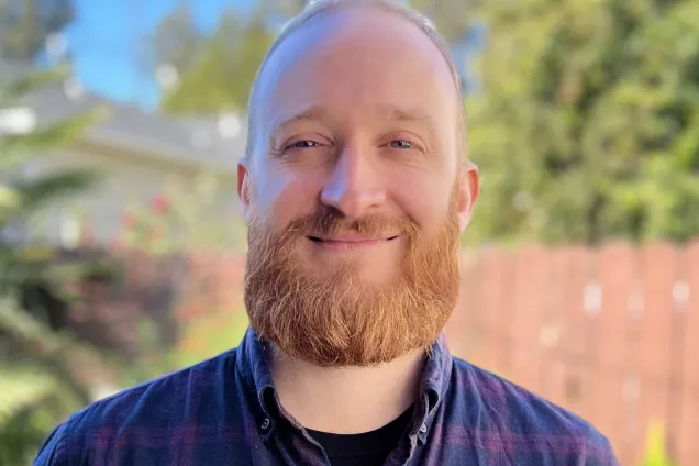
Dr. Jacob Vogel
DDLS Fellow, Lund University
Leveraging disease progression models toward biological insights and clinical practice in neurodegenerative disease
Neurodegenerative diseases are characterized by accumulation of pathological proteins leading to progressive damage to select neuronal populations and concomitant clinical decline. Models have emerged that build around biological properties of these diseases in order to model their progression. Many of these “disease progression models” were initially employed to provide holistic descriptions of the disease process, or to test specific hypotheses. However, insofar as disease progression models act as multifaceted simulations of a disease, they have great potential to be extended to provide novel biological insights and even clinically useful information. In this talk, I will discuss work using two different types of disease progression models. I will first discuss connectome-based models — which operate under the assumption that disease pathology travels through the brain’s intrinsic axonal architecture — in the context of accumulation of the tau protein in Alzheimer’s disease. Second, I will discuss applications of the Subtype and Stage Inference disease subtyping algorithm across a different neurodegenerative diseases, including Alzheimer’s disease, Lewy body disease and TDP-43 proteinopathy. Throughout the talk, there will be a specific focus on how we and others have employed these models to formulate new hypotheses about disease biology, as well as preliminary work toward building disease progression models into clinically useful tools.

Dr. Farouk Dako
Assistant Professor of Radiology, University of Pennsylvania
Talk title: TBD
TBD
Agenda
| Time | Session |
|---|---|
| 1:30 PM - 1:35 PM | Welcome and introduction |
| 1:35 AM - 2:20 PM |
Keynote 1
Dr. Jacob Vogel |
| 2:20 PM - 2:31 PM |
DP-MoSt for detecting sub-trajectories in the natural history of pathologies
Alessandro Viani |
| 2:31 PM - 2:42 PM |
Back to the Future: Challenges of Sparse and Irregular Medical Image Time Series
Nico A Disch |
| 2:42 PM - 2:53 PM |
Probabilistic Temporal Prediction of Continuous Disease Trajectories and Treatment Effects Using Neural SDEs
Joshua D Durso-Finley |
| 2:53 PM - 3:04 PM |
Enhancing Spatiotemporal Disease Progression Models via Latent Diffusion and Prior Knowledge
Lemuel Puglisi |
| 3:04 PM - 3:15 PM |
Individualized multi-horizon MRI trajectory prediction for Alzheimer's Disease
Rosemary Y He |
| 3:15 PM - 3:30 PM |
Towards Longitudinal Characterization of Multiple Sclerosis Atrophy Employing SynthSeg Framework and Normative Modeling
Pedro Macias Gordaliza |
| 3:30 PM - 4:00 PM | Coffee Break |
| 4:00 PM - 4:45 PM |
Keynote 2
Dr. Farouk Dako |
| 4:45 PM - 4:50 PM |
Automated Segmentation and Registration of Fundus Autofluorescence Imaging facilitates better monitoring of Geographic Atrophy Growth
Yoga Advaith Veturi |
| 4:50 PM - 5:01 PM |
SegHeD: Segmentation of Heterogeneous Data for Multiple Sclerosis Lesions with Anatomical Constraints
Berke D Basaran |
| 5:01 PM - 5:12 PM |
Longitudinal Segmentation of MS Lesions via Temporal Difference Weighting
Maximilian R Rokuss |
| 5:12 PM - 5:23 PM |
Registration of Longitudinal Liver Examinations for Tumor Progress Assessment
Walid Yassine |
| 5:23 PM - 5:34 PM |
Tracking lesion evolution using a Boundary Enhanced Approach for MS change segmentation (BEAMS)
Prateek Mathur |
| 5:34 PM - 5:45 PM |
A Radiological-based Coordinate System for the Human Body: A Proof-of-Concept
Stefano Trebeschi |
| 5:45 PM - 5:56 PM |
Vestibular schwannoma growth prediction from longitudinal MRI by time conditioned neural fields
Yunjie Chen |
| 5:56 PM - 6:00 PM | Closing remarks and adjournment |
Organizers
and Fraunhofer MEVIS, Germany
University College London, London, UK
University College London, London, UK

Portland State University, Portland, USA

University College London, London, UK

Lund University, Lund, Sweden

University College London, London, UK
University of Sussex, Brighton, UK

Clinical Center, Bethesda, USA
IRCCS Ospedale Policlinico San Martino, Genoa, Italy

Lübeck, Germany,
Clinical Center, Bethesda, USA

University College London, London, UK

